
Key Takeaways
- Prepare the patient, room, equipment, and team roles before each procedure.
- Monitor fluid use and losses continuously and communicate changes promptly.
- Use clear communication and defined responsibilities throughout the case.
- Match tissue-removal techniques and equipment to the patient and procedure.
- Use checklists and case reviews to identify recurring delays or workflow gaps.
- Provide clear discharge instructions and follow-up guidance to support patient care.
Operative hysteroscopy depends on much more than the procedure itself. The most reliable cases begin with a shared plan, a prepared room, clear responsibilities, and an approach that keeps patient safety at the center of every decision. Whether the team is treating a polyp or a submucosal fibroid, a consistent workflow can reduce preventable interruptions and support better coordination. For gynecology teams, efficiency should never mean rushing. It means removing uncertainty before it affects the case, communicating clearly when conditions change, and giving patients understandable guidance before discharge. A dependable process also helps physicians, nurses, anesthesia professionals, surgical technologists, and administrative staff work from the same expectations.
Why Workflow Matters in Operative Hysteroscopy
Small process gaps can create larger clinical problems. A missing supply, an unclear fluid-monitoring assignment, an unlabeled specimen container, or an equipment issue can disrupt concentration at an important moment. A standard workflow provides a dependable foundation while leaving room for clinical judgment when anatomy, lesion characteristics, patient health, or procedural complexity require a different plan. Teams should define efficiency as reliable preparation and fewer avoidable handoffs, not as a shortcut around safety checks. Current guidance on hysteroscopy safety emphasizes appropriate patient selection, staff competency, equipment readiness, and awareness of complications associated with distending media.
Steps to Complete Before the Procedure
A brief pre-procedure review helps the team enter the room with the same understanding of the case. The review should be concise enough to use consistently and detailed enough to identify meaningful risks.
- Confirm patient identity, planned procedure, consent, allergies, and relevant medical history.
- Review imaging, prior uterine procedures, medications, bleeding concerns, and anesthesia considerations.
- Discuss the intended treatment, anticipated findings, and circumstances that could require a pause, staged treatment, or change in approach.
- Verify the availability of visualization equipment, irrigation fluid, sterile instruments, energy equipment, and specimen supplies.
- Assign responsibility for fluid tracking, specimen handling, documentation, and patient updates.
Building a Clear Room Setup
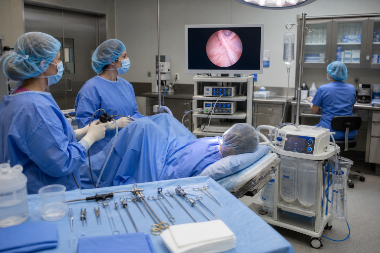
A consistent room layout improves situational awareness. Position the monitor where the surgeon and essential staff can view it comfortably. Organize cables, tubing, foot controls, suction, and irrigation lines before the patient enters the room. Complete required equipment checks and prepare labeled specimen containers before tissue removal begins. Standardization does not require every case to look identical. It means staff know where to find essential items and understand how the setup should change for a more complex procedure. This familiarity can support smoother turnover and reduce time spent searching for supplies after the case has started.
Fluid Monitoring and Patient Safety
Fluid management deserves continuous attention during operative hysteroscopy. The team should understand the planned distention medium, monitor inflow and outflow in real time, calculate the fluid deficit per facility policy, and communicate the total at agreed-upon intervals. Waiting until the end of the case to reconstruct measurements from memory introduces unnecessary risk. One trained person should have explicit responsibility for tracking and documenting fluid totals, but the surgeon and anesthesia team should remain aware of any changes in values. Teams may establish more conservative stopping thresholds for patients with conditions that could increase the consequences of fluid absorption or overload. Changes in breathing, oxygenation, mental status, nausea, chest discomfort, or vital signs require prompt assessment and a coordinated decision about whether to pause or end the procedure.
Team Communication During the Case
Short, direct communication helps the room respond before a concern becomes an emergency. Before instrumentation begins, confirm the procedure, expected pathology, key risks, and potential stopping points. During the case, call out changes in visualization, bleeding, fluid balance, equipment performance, or patient condition. Closed-loop communication is especially valuable for urgent requests. The receiver repeats the request, confirms completion, and reduces the chance that a critical instruction is missed. Before the patient leaves the room, confirm specimen labels, destinations, documentation, and the immediate recovery plan.
Planning Tissue Removal with Care
Review the lesion’s location, size, depth, and number before treatment begins. The selected approach should match the patient’s needs, available equipment, and the clinician’s training and experience. Throughout removal, maintain visualization, address bleeding when it compromises the operative view, and pause to reassess whenever anatomy becomes unclear or expected progress stalls. Device selection remains an individualized clinical decision. In a 2026 prospective, single-center observational study, smaller-diameter operative hysteroscopes were associated with fewer intraoperative complications, while completion rates and operating times were similar. That association is useful for discussion, but it does not establish one device choice as appropriate for every patient or procedure.
Ways to Reduce Delays and Rework
- Use one visible setup guide. Keep it practical, brief, and easy to revise when processes change.
- Prepare specimens early. Have correct containers and labels ready before removal begins.
- Inspect equipment between cases. Replace missing, expired, or damaged items before the next patient arrives.
- Track repeat causes of delay. Look for patterns involving supplies, equipment checks, documentation, or unclear orders.
- Create a backup plan. Discuss responses to poor visualization, increased bleeding, equipment failure, or an incomplete procedure.
Supporting the Patient Before and After Surgery
A strong workflow includes the patient experience. Use plain language to explain why the procedure is being performed, what recovery may involve, and how the team will communicate findings. Written discharge instructions should address medications, expected cramping or spotting, activity guidance, follow-up, and symptoms that should prompt a call to the care team.
Patients should also have a practical way to ask questions after discharge. Clear expectations can reduce anxiety and help patients recognize when they need timely medical advice.
Using Checklists and Case Reviews
Quality improvement does not need to create burdensome paperwork. Review a small number of useful measures, such as setup delays, cancellations, incomplete procedures, fluid-documentation gaps, specimen errors, and recurring equipment problems. Discuss complications in a learning-focused setting, compare actual practice with the written protocol, and ask staff where the workflow feels unclear or repetitive.
Common Questions About Operative Hysteroscopy
What is the main goal of a workflow?
The goal is safe, coordinated care from preparation through recovery, with clear roles, reliable monitoring, and appropriate clinical decision-making.
When should a procedure be paused?
A pause may be appropriate when visualization is poor, fluid limits are reached, bleeding cannot be adequately managed, equipment fails, or the patient’s condition changes.
Can one checklist fit every case?
A checklist provides a reliable baseline, but it cannot replace clinical judgment. Complex pathology, unusual anatomy, and higher-risk patients may require additional planning.
Conclusion
A better operative hysteroscopy workflow begins before the procedure and continues through follow-up. Careful preparation, organized equipment setup, appropriate patient assessment, real-time fluid awareness, concise communication, and clear role assignments can help the team maintain an orderly process. During the procedure, monitoring fluid use and losses, documenting important events, and responding promptly to changes are important parts of safe practice. Afterward, accurate documentation, appropriate recovery monitoring, specimen handling when applicable, and clear follow-up instructions help maintain continuity of care. Reviewing each stage can also help teams identify workflow gaps and opportunities for improvement. By combining preparation, communication, monitoring, documentation, and post-procedure review, healthcare teams can reduce avoidable disruption while keeping patient safety and quality of care central throughout the process.
photo by depositphotos